

Melanoma
Melanoma, also known as malignant melanoma, is a type of skin cancer that arises from melanocytes. It is the most aggressive common skin cancer and has the highest risk of metastasis compared with BCC and cutaneous SCC.
This updated UKMLA guide to melanoma is based on NICE NG14 and PCDS guidance, which covers causes, risk factors, recognition, diagnosis, and management.
Causes and Risk Factors
Melanoma is thought to arise from uncontrolled growth of melanocytic stem cells that have undergone genetic mutations:
- ~90% of mutations are sporadic (most common: BRAF V600E mutation [Ref])
- ~10% of mutations are inherited (most common: CDKN2A mutation)
Risk factors:
| High-risk (10x risk) | Patients with:
|
| Moderate-risk (6-10x risk) |
|
| Slightly increased risk (1-3x risk) |
|
Most (~80%) melanomas arise de novo, the remaining arise from pre-existing naevi.
Clinical Features
Shared Features
The ABCDE rule is often used to remember the red flag features of melanoma:
| Letter | Feature | Description |
|---|---|---|
| A | Asymmetry | One half of the lesion does not match the other half in shape or appearance (e.g. by drawing a line through the middle of the lesion) |
| B | Border | Irregular / poorly defined border |
| C | Colour | Multiple colours / uneven pigmentation (e.g. black, brown, blue, red, white, pink) |
| D | Diameter | Diameter >6 mm |
| E | Evolving | Any change in size / shape / colour / elevation / symptoms (e.g. itching, bleeding, crusting) |
Type-Specific Features
| Type | Description / key features |
|---|---|
| Superficial spreading melanoma | Most common subtype
Flat lesion with the classic ABCDE features mentioned above |
| Nodular melanoma | Most aggressive subtype; more common in 50-60 y/o males
Classically lacks the ABCDE features, but presents with EFG:
|
| Lentigo maligna melanoma | 2nd most common subtype; it occurs around hair follicles – most commonly the head and neck
Peak incidence: 65-80 y/o Flat lesion with the classic ABCDE features mentioned above |
| Acral lentiginous melanoma | Least common subtype (2-3%), it is NOT related to UV exposure
Typically affects:
Most common type of melanoma in people with coloured skin types |
Click to See Clinical Images
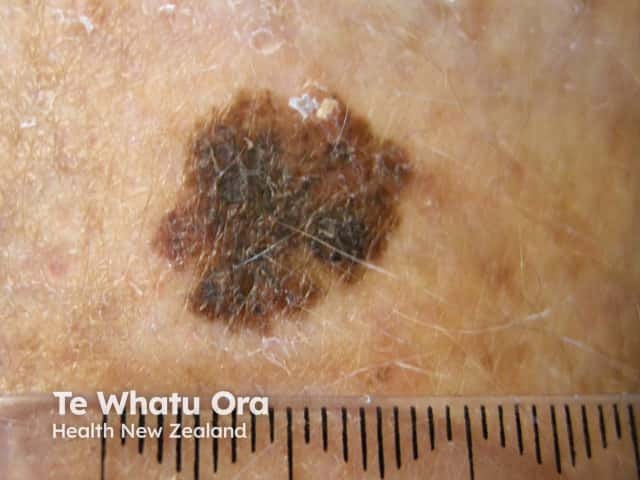
Superficial spreading melanoma
Source: https://dermnetnz.org/topics/superficial-spreading-melanoma
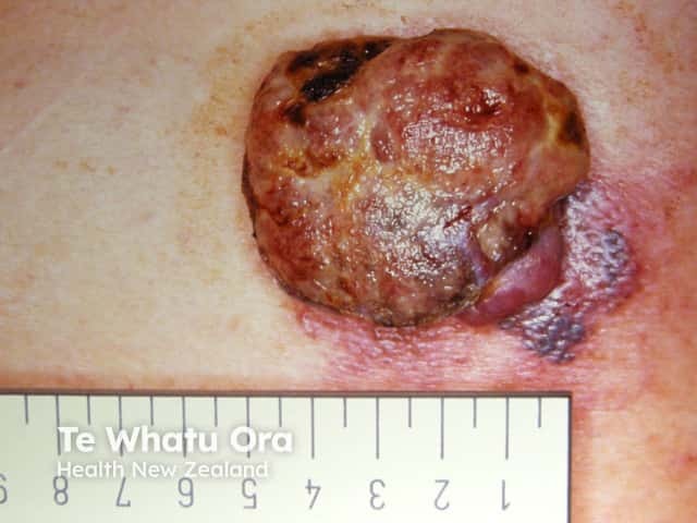
Nodular melanoma
Source: https://dermnetnz.org/topics/nodular-melanoma
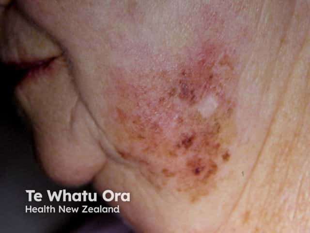
Lentigo maligna
Source: https://dermnetnz.org/topics/lentigo-maligna-and-lentigo-maligna-melanoma
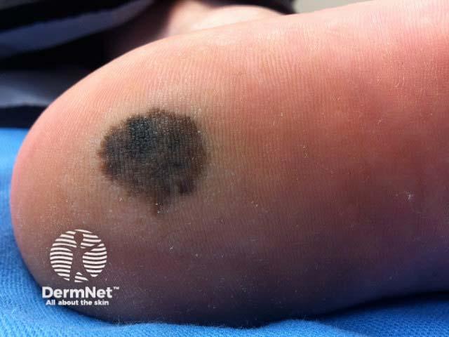
Acral lentiginous melanoma on the heel
Source: https://dermnetnz.org/topics/acral-lentiginous-melanoma
Complications
Melanoma is the most likely skin cancer to metastasise, accounting for the majority of skin cancer-related mortality. [Ref]
Most common sites of distant metastasis: lung, liver, bone, lymph nodes. [Ref]
Red Flags and Referral Criteria
Suspected cancer pathway referral if there is:
- A pigmented skin lesion with ≥3 on weighted 7-point checklist score, or
- Dermoscopy suggests melanoma
- Nodular melanoma suspected (consider)
Weighted 7-point checklist:
- Major features (2 points each)
- Change in size
- Irregular shape
- Irregular colour
- Minor features (1 point each)
- ≥7mm (largest diameter)
- Inflammation
- Oozing
- Change in sensation
Investigation and Diagnosis
Gold standard: full-thickness excisional biopsy
- To assess Breslow depth, the single most important prognosis predictor
- Avoid incisional biopsy if possible, as it will prevent accurate staging
Staging:
- Sentinel lymph node biopsy
- Do not offer in stage IA melanoma
- Consider if Breslow thickness >1.0 mm or 0.8-1.0 mm + ulceration / lymphovascular invasion / mitotic index ≥2
- Cross-sectional imaging (to check for distant metastasis in advanced disease)
- Whole body and brain contrast-enhanced CT / MRI
- Consider / offer in stage IIB and onwards
Other investigations:
- Vitamin D level
- BRAF mutation analysis – do not offer in stage IA / IB (only in stage IIA and onwards)
TNM Staging
The main purpose of including TNM staging is to provide context for NICE’s management recommendations, which are guided by tumour stage.
| Stage | T (tumour) | N (node) | M (metastasis) |
|---|---|---|---|
| 0 |
|
-ve | -ve |
| I |
|
||
| II |
|
||
| III |
|
+ve | |
| IV | +ve / -ve | +ve |
Management
Definitive management: surgical excision
Specifically, what happens in melanoma:
- Initially, an excisional biopsy is performed to diagnose melanoma and assess the Breslow depth
- After melanoma is diagnosed, a wide local excision is performed (where a further excision is performed around the previous biopsy site with a wider safety margin to reduce the risk of local recurrence)
Breslow Depth-Guided Management
Breslow depth-guided wide local excision margin.
| Breslow depth | Wide local excision margin |
|---|---|
| Melanoma in situ | 0.5–1 cm |
| ≤1 mm | 1 cm |
| 1.1-2.0 mm | 1–2 cm |
| >2 mm | 2 cm |
Disclaimer:
Note that this is no longer explicitly mentioned in the 2022 NICE guideline, but it was included in the 2015 version and reflects traditional teaching and standard clinical practice.
Also see the TNM stage-guided management below, which is in line with the NICE 2022 guideline.
TNM Stage-Guided Management
The latest NICE guideline recommends that management should be guided by TNM stage (see above), rather than Breslow depth alone (NB Breslow depth plays a role in influencing the TNM stage):
| TNM stage | Recommended management |
|---|---|
| 0 | 1st line: wide local excision with surgical margin of 0.5 cm
Consider topical imiquimod + repeat skin biopsy if surgery would lead to unacceptable disfigurement or morbidity. |
| I-II | 1st line: wide local excision with surgical margin of:
|
| III | 1st line: wide local excision
Other therapy:
If unresectable: offer systemic therapy (see below) |
| IV | 1st line: systemic therapy
If immunotherapy and targeted therapy are not appropriate → consider chemotherapy with dacarbazine |
References
Related Articles
Skin Cancer – Recognition and Referral