

Benign Skin Lesions
Benign skin lesions are non-cancerous skin growths that are commonly encountered in clinical practice. This article covers high-yield benign skin lesions relevant to UKMLA-level dermatology based on PCDS guidance, including melanocytic naevi, seborrhoeic keratosis, dermatofibroma, epidermoid cysts, and pyogenic granuloma.
Melanocytic Naevi
Definition
Melanocytic naevi, commonly known as moles, are benign proliferations of melanocytes within the skin.
The cause of melanocytic naevi is unknown
Note on terminology:
- A single lesion is called a melanocytic naevus
- Multiple lesions are called melanocytic naevi
Clinical Features
Most melanocytic naevi become apparent during childhood or early adult life
Benign Melanocytic Naevia
Benign melanocytic naevi are the most common type of melanocytic naevi
- Symmetrical shape
- Smooth, regular border
- Uniform colour (usually brown)
- Flat or slightly raised surface
- Stable appearance over time
- May lighten with age
Atypical (Dysplastic) Melanocytic Naevi
Atypical melanocytic naevi are still benign, but they may appear irregular and can overlap clinically with malignant melanoma. Their main implication is that people with multiple atypical naevi have a higher overall risk of malignant melanoma.
Features of atypical (dysplastic) melanocytic naevi:
- Large (often >7 mm)
- Irregular borders are common
- Colour varies (uniform colour or >1 colour)
Some specific syndromes:
- Atypical mole syndrome: presence of >50 melanocytic naevi with at least 3 atypical melanocytic naevi
- Familial atypical mole and melanoma syndrome (FAMMM): presence of >50 typical and atypical melanocytic naevi PLUS a family history of melanoma
- 20-40% of these individuals have a mutation in the CDKN2A gene
- They are 25x more likely to develop malignant melanoma
Malignant Melanoma Red Flags
The most important differential of melanocytic naevi is malignant melanoma.
Red flags suggesting malignant melanoma – ABCDE rule:
- A – Asymmetry
- B – border irregular / poorly defined
- C – colour more than 1
- D – diameter >6 mm
- E – evolving (change in size / shape / colour)
Ugly duckling sign: A pigmented lesion should be considered suspicious if it looks noticeably different from the patient’s other naevi, even if it does not clearly meet all ABCDE criteria.
Click to See Clinical Images
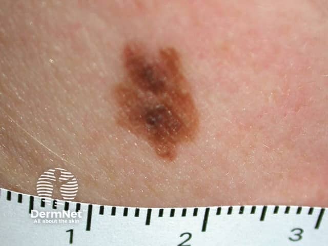
Small congenital melanocytic naevus
Source: https://dermnetnz.org/topics/melanocytic-naevus
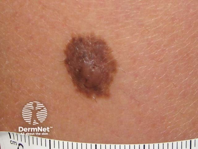
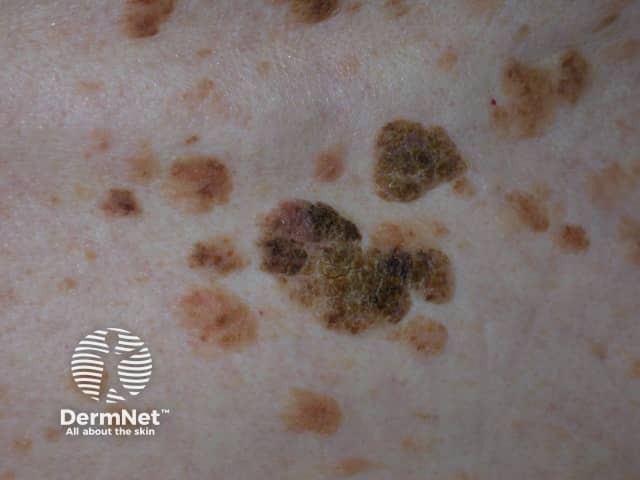
Atypical melanocytic naevus
Source: https://dermnetnz.org/topics/atypical-melanocytic-naevus
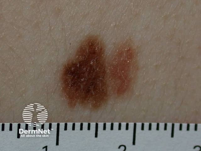
Atypical melanocytic naevus
Source: https://dermnetnz.org/topics/atypical-melanocytic-naevus
Management
Benign Melanocytic Naevi
No specific treatment is necessary, as the chance of any one individual naevus becoming malignant is very low
Provide general advice to ALL patients:
- The larger the number of naevi = the greater the risk of melanoma
- Patients with >100 naevi on their body have a ~7x higher risk of developing melanoma, compared to those with ≤15 naevi
- Having ≥11 naevi on an arm predicts an increased risk of having >100 total body naevi
- Educate on self-examination of moles (the ABCDE rule mentioned above is helpful)
Atypical (Dysplastic) Melanocytic Naevi
Provide general advice to ALL patients:
- The larger the number of naevi = the greater the risk of melanoma
- Patients with >100 naevi on their body have a ~7x higher risk of developing melanoma, compared to those with ≤15 naevi
- Having ≥11 naevi on an arm predicts an increased risk of having >100 total body naevi
- Educate on self-examination of moles (the ABCDE rule mentioned above is helpful)
Patient should take photographs of their moles for reference and future comparison
Referral indications:
- Patients with only a few atypical melanocytic naevi and without a family history of malignant melanoma do NOT need referral
- Routine referral is indicated if
- Presence of a large number of naevi – mixture of typical and atypical melanocytic naevi (for thorough dermoscopic assessment and photography)
- Familial atypical mole and melanoma syndrome (FAMMM): presence of >50 typical and atypical melanocytic naevi PLUS a family history of melanoma
References
Seborrhoeic Keratosis
Definition
Seborrhoeic keratosis is a benign overgrowth of epidermal keratinocytes of the skin.
The cause of seborrhoeic keratosis is unknown.
Seborrhoeic keratosis is also known as seborrhoeic wart or basal cell papilloma (NOT basal cell carcinoma!).
Clinical Features
Most commonly present in middle-aged and older individuals (>40 y/o)
Symptoms:
- Usually asymptomatic
- Itching is possible
- Non-tender
Signs:
- Location: face and trunk most common
- Colour: brown / black / yellow
- Size: most common 1-3 cm, but can be very large
- Appearance
- Scaly / greasy appearance +/- irregular verrucous surface
- ‘Stuck-on’ appearance – lesion sits on normal skin, thus giving the impression it is ‘stuck on’ the skin and can be easily peeled off
Leser-Trélat sign: Sudden eruption of multiple seborrhoeic keratoses with rapid increase in size and number.
This is traditionally taught as a paraneoplastic syndrome, associated with gastric or colorectal cancer
- This remains highly debated
- However, if one presents with the Leser-Trélat sign PLUS other red flag symptoms (e.g. weight loss, change in bowel habits), the patient should be urgently referred for further assessment
Despite the similar name, seborrhoeic keratosis is completely different from seborrhoeic dermatitis – a chronic inflammatory skin condition causing itchy, greasy, red or flaky patches.
Click to See Clinical Images
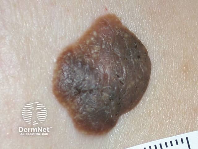
Seborrhoeic keratosis
Source: https://dermnetnz.org/topics/seborrhoeic-keratosis
Seborrhoeic keratosis
Source: https://dermnetnz.org/topics/seborrhoeic-keratosis
Management
Most seborrhoeic keratoses do NOT require treatment
If removal is necessary (e.g. uncertain diagnosis, symptomatic, possible malignancy, cosmetic concern):
- Most common: cryotherapy with liquid nitrogen
- Very thick lesions can be removed by curettage and cautery
- All samples should be sent for histology
References
Dermatofibroma
Definition
Dermatofibroma is a common benign fibrous raised skin lesion.
The cause of dermatofibroma is unknown.
Dermatofibroma is also known as histiocytoma.
Clinical Features
Dermatofibroma can arise after an insect bite or local trauma
- More common in females
- Young adults are most commonly affected
Symptoms:
- Usually asymptomatic
- Can be painful when knocked against
Signs:
- Location: lower limbs (esp. lower legs and thigh)
- Colour: skin coloured / brown / black
- Size: most common 5 mm
- Appearance
- Slightly elevated (papule)
- More pigmented at the periphery (edges)
- On palpation (important for diagnosis)
- Dimple sign (positive pinch sign): lateral compression (pinching) causes a central dimple in the lesion
- Palpable “iceberg” component: the lesion may feel larger than it appears visually, with a regular, firm, rubbery dermal lump palpable beneath the surface
Click to See Clinical Images
Dermatofibroma
Source: https://dermnetnz.org/topics/dermatofibroma
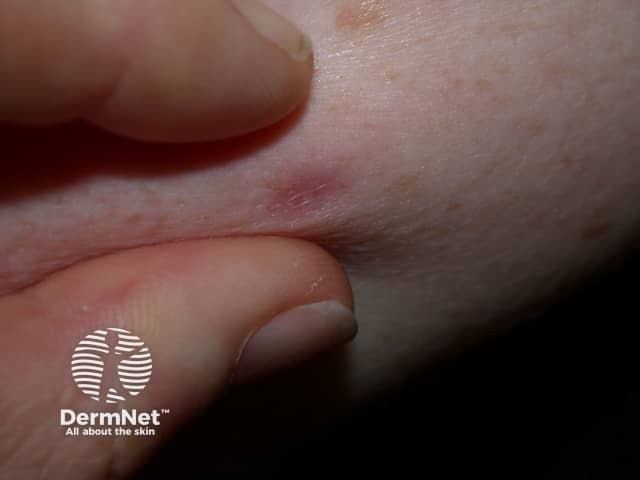
Dimple sign seen in dermatofibromas
Source: https://dermnetnz.org/topics/dermatofibroma
Management
Dermatofibromas do not normally require treatment because they are benign lesions with no malignant potential.
References
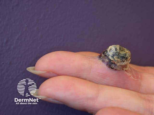
Epidermoid Cyst
Definition
An epidermoid cyst is a cutaneous cyst that contains keratin and its breakdown products, surrounded by an epidermoid wall.
Clinical Features
Most commonly affects young and middle-aged adults
Symptoms:
- Asymptomatic
- Unless infected
Signs:
- Location: face, neck, shoulder and chest
- Colour: yellow-white
- Slow-growing
- Appearance
- Firm, elastic, dome-shaped nodule
- +/- Central punctum (black-coloured keratin)
Infected cysts enlarge, becoming red and tender, and eventually discharge pus.
Click to See Clinical Images
Epidermoid cyst
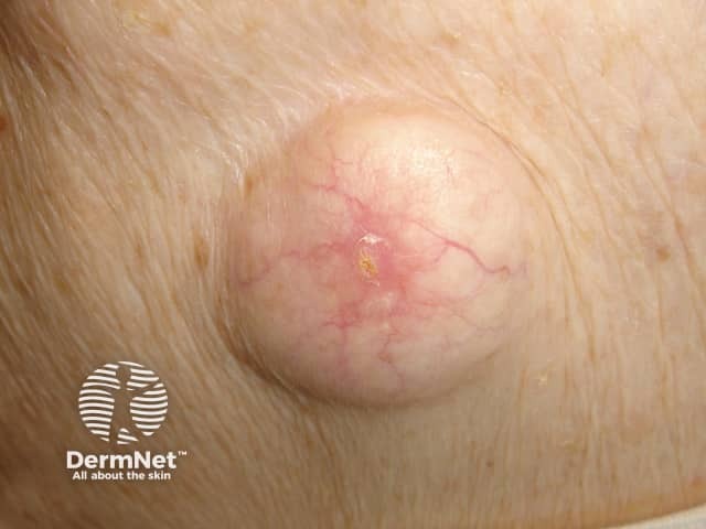
Source: https://dermnetnz.org/topics/epidermoid-cyst
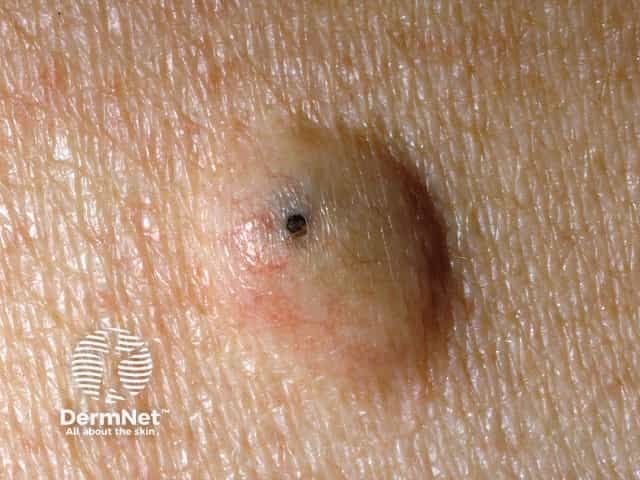
Epidermoid cyst with characteristic punctum
Source: https://dermnetnz.org/topics/epidermoid-cyst
Management
Most uncomplicated asymptomatic epidermoid cysts do not require treatment
- Surgical excision is the most effective treatment (e.g. for cosmetic reasons, bothersome symptoms)
- For an infected cyst: initial antibiotics followed by incision and drainage after the infection settles
References
Pyogenic Granuloma
Definition
Pyogenic granuloma is a benign, rapidly growing vascular lesion.
Clinical Features
Most commonly occur in children, young adults and during pregnancy
Often appears after trauma to the skin area
- Location: fingers, hands, head and upper trunk most common
- Rapid growing – starts as a small red spot that quickly grows into a nodule
- Initially smooth, but then becomes eroded and bleeds significantly on contact / after minimal trauma
Click to See Clinical Images
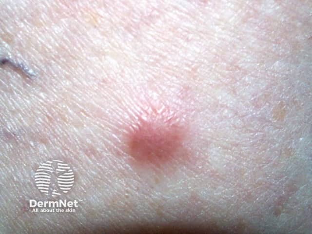
Pyogenic granuloma
Source: https://dermnetnz.org/topics/pyogenic-granuloma
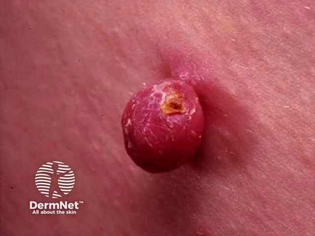
Pyogenic granuloma
Source: https://dermnetnz.org/topics/pyogenic-granuloma
Management
Apply Vaseline to the surrounding skin, then apply as much table salt as possible to the whole lesion, then cover with a dressing (e.g. surgical tape or Clingfilm)
Repeat the above process every day until the lesion resolves.
References
Related Articles
Skin Cancer – Recognition and Referral