

Cutaneous Squamous Cell Carcinoma (cSCC)
Cutaneous squamous cell carcinoma (cSCC) is the 2nd common type of skin cancer, arising from keratinocytes of the epidermis.
This updated UKMLA guide to cutaneous squamous cell carcinoma (cSCC) is based on BAD, NICE, and PCDS guidance, which covers risk factors, clinical features, referral criteria, diagnosis and management.
Causes and Risk Factors
Key risk factors:
- Cumulative lifetime UV exposure – major risk factor
- Sunlight
- Sunbeds
- Artificial UV exposure (e.g. psoriasis management)
- Fair-skin type (type 1-2)
- Advancing age
- Males
Importantly, individuals who are immunosuppressed (esp. those after organ transplantation) are at a much higher risk of developing cSCC.
Clinical Features
| Location | Most common on sun-exposed sites:
|
| Appearance |
|
cSCC variants:
- Keratoacanthoma: a low-grade variant of SCC, covered in the Pre-Malignant Skin Lesions article
- Marjolin ulcer: aggressive SCC arising from a scar or inflammation
Symptoms
- The presence of pain or tenderness should raise suspicion for SCC.
- SCC is more frequently painful than BCC or melanoma (SCC > BCC > melanoma), which are typically asymptomatic.[Ref]
Click to See Clinical Images
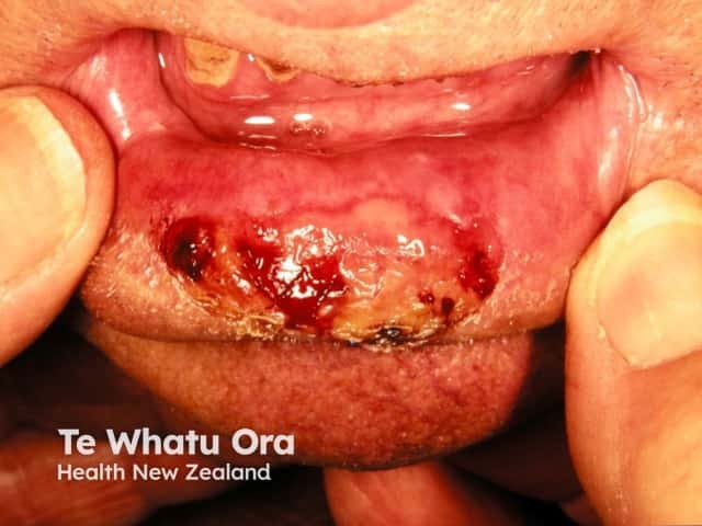
Squamous cell carcinoma
Source: https://dermnetnz.org/topics/cutaneous-squamous-cell-carcinoma
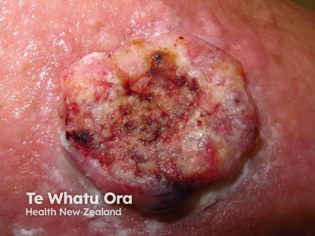
Squamous cell carcinoma
Source: https://dermnetnz.org/topics/cutaneous-squamous-cell-carcinoma
Complications
cSCC carries the risk of metastasis; the risk is higher in immunosuppressed individuals
Most common location: lymph nodes
Red Flags and When to Refer
Consider a suspected cancer pathway referral if there is a suspected squamous cell carcinoma skin lesion.
Investigation and Diagnosis
Gold standard: skin biopsy for histology
- Preferred: full-thickness incisional biopsy (containing both peripheral and deep margins)
- Large / anatomically challenging area → incisional (punch) biopsy
Management
Consider Mohs micrographic surgery if:
- Tumour margins are not clearly visible / well-defined
- At sites where tissue conservation is important (e.g. eyelid, lips, ears, fingers, genitalia)
- At cosmetically sensitive areas (e.g face)
If surgery is not appropriate: primary radiotherapy is an option
Surgical Margins
Peripheral surgical margins (determined under magnification / dermoscopy):
- Low risk tumour → ≥4 mm margin
- High risk tumour → ≥6 mm margin
- Very high risk tumour → ≥ 10mm margin
Also ensure at least 1 mm histological clearance at all margins.
Tumour risk definitions:
| Low Risk | High Risk | Very High Risk |
|---|---|---|
| ALL the following must be met | ANY of the following | ANY of the following |
|
|
|