

Ischaemic Stroke
Ischaemic stroke accounts for ~85% of stroke cases, it occurs when cerebral blood flow is interrupted, causing brain ischaemia and infarction.
This updated UKMLA guide to ischaemic stroke is based on National Clinical Guideline for Stroke and NICE NG128, which covers guidelines on initial assessment, diagnosis, imaging, reperfusion therapy including thrombolysis and thrombectomy, and long-term management.
Scope of article:
- This article focuses on the initial assessment and management of stroke, with particular emphasis on ischaemic stroke.
- General background information, including stroke types, causes, stroke mimics and vascular territory symptoms, is covered in the Stroke (Overview) article.
- Specific management of haemorrhagic stroke is covered separately in the Haemorrhagic Stroke article.
Approach (All Strokes)
Refer all suspected cases of stroke to a hyperacute stroke service
The pathway below illustrates a simplified approach to acute stroke assessment and initial management in the emergency department / hospital setting.
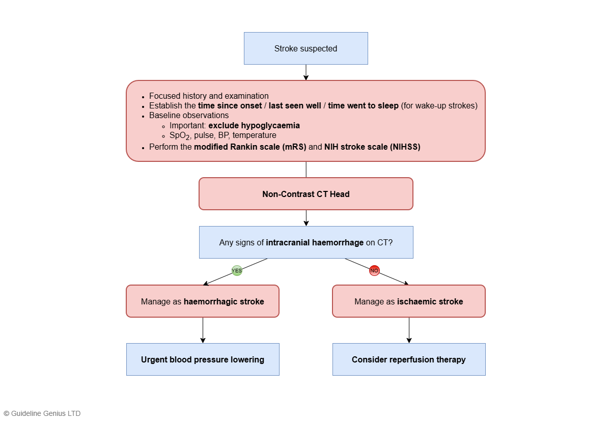
Assessment and Work-Up
Refer all suspected cases of stroke to a hyperacute stroke service.
1. Initial Clinical Assessment
- Exclude hypoglycaemia with a capillary blood glucose test
- Clinical history and assessment, including
- Establish time of onset / last seen well / time went to sleep (for wake-up stroke)
- If possible, perform the NIHSS and mRS
- Check for any thrombolysis contraindications
- Clinical observations (e.g. SpO2, pulse, blood pressure)
- Screen swallowing before the patient receives anything by mouth
2. Brain Imaging
1st line and most important: non-contrast CT head within 1 hour to exclude intracranial haemorrhage
Further imaging:
- If there is diagnostic uncertainty → MRI brain with stroke-specific sequences
- To assess eligibility for thrombolysis and/or thrombectomy
- CT angiogram from the aortic arch to skull vertex – often added to the initial CT if the patient is potentially eligible
- Perfusion imaging: CT / MR perfusion (alternative: MRI measuring DWI-FLAIR mismatch) – for delayed presentation
In practice, if the patient is likely eligible for thrombectomy (e.g. high NIHSS and low mRS), a CT stroke protocol is usually ordered instead of just a non-contrast CT head.
A non-contrast CT head is still the most important initial imaging; it is just performed as part of the CT stroke protocol.
CT stroke protocol includes a sequence of scans: non-contrast CT → CT perfusion → CT angiography, which excludes intracerebral haemorrhage and gives the relevant information needed to assess eligibility for thrombectomy.
3. Imaging Interpretation and Next Steps
As part of the acute stroke work-up, the main purpose of a non-contrast CT head is to identify or exclude intracranial haemorrhage, indicated by
- A hyperdense (bright-white) area of acute blood
- May be surrounded by a hypodense rim of oedema
- +/- Mass effect (e.g. midline shift, ventricular compression)
Subsequent management:
- If CT shows intracranial haemorrhage → manage as haemorrhagic stroke (covered in the Haemorrhagic Stroke article)
- If CT shows no haemorrhage → manage as ischaemic stroke (see below)
- If the clinical presentation and/or CT suggests subarachnoid haemorrhage → see the Subarachnoid haemorrhage (SAH) article
The initial non-contrast CT head in suspected stroke is NOT performed to confirm ischaemia, but to exclude haemorrhage.
If the clinical presentation is consistent with an ischaemic stroke and the CT does not show haemorrhage, then the patient is treated as having an ischaemic stroke.
This is because CT head is often normal in the first few hours after an ischaemic stroke; visible hypodensity may not appear until 12–24 hours after onset. An MRI can detect ischaemic changes within minutes of ischaemia, but should not delay urgent management.
Imaging Findings in Ischaemic Stroke
Imaging findings of ischaemic stroke:
| CT head (non-contrast) |
|
| MRI |
MRI can detect very early ischaemic changes (within minutes) |
On CT/MRI, the location of the infarct (hypodensity on CT / hyperintensity on MRI) reflects the arterial territory affected:
- Lateral cortex (away from midline) → MCA infarct
- Medial cortex (near the midline) → ACA infarct
- Occipital lobe → PCA infarct
- Basal ganglia / internal capsule → lenticulostriate vessel infarct (Lacunar stroke)
- Brainstem / cerebellum → vertebrobasilar artery infarct
Acute Management
Supportive Measures / General Management
| Oxygen therapy | Do not give oxygen routinely, unless SpO2 <94% |
| Blood glucose | Target: 4-11 mmol/L |
| Swallowing assessment | Screen swallowing before any oral intake (food, fluid, medications)
|
| Blood pressure control | Blood pressure should NOT be routinely lowered in acute ischaemic stroke unless there are specific indications, as this can further compromise cerebral perfusion & neurological outcomes
Indications to lower the blood pressure:
|
Aspirin
Aspirin 300 mg should be offered ASAP once
- Intracranial haemorrhage has been excluded on the initial non-contrast CT, and
- Patient is NOT undergoing thrombolysis (including those who are only undergoing thrombectomy)
If the patient is dysphagic → administer via an enteral / rectal tube
If the decision is made to treat the patient with thrombolysis, aspirin 300mg should only be given 24 hours AFTER thrombolysis, once repeat imaging excludes haemorrhage.
Do NOT give aspirin before or with thrombolysis.
Reperfusion Therapy
Reperfusion therapy aims to restore blood flow to ischaemic brain tissue. In acute ischaemic stroke, this may involve:
- Thrombolysis, which dissolves the clot using a clot-busting drug, and/or
- Thrombectomy, which mechanically removes a clot from a large artery
Thrombolysis and thrombectomy are 2 independent interventions, with separate indications and contraindications:
- If the patient is eligible for both thrombolysis and thrombectomy → both should be arranged urgently, without either treatment delaying the other
- Do NOT skip thrombolysis to facilitate a thrombectomy (if eligible for both)
- Do NOT delay thrombectomy for observation after thrombolysis
Thrombolysis
Thrombolysis involves administering a “clot-busting drug” to dissolve an occluding thrombus and restore blood flow in acute ischaemic stroke
- Agent of choice: IV alteplase / tenecteplase
- Before undergoing thrombolysis, the patient must be 1) eligible and 2) have no contraindications
Thrombolysis should not be delayed for advanced imaging.
Advanced imaging, such as CT angiography, may be required to confirm eligibility and plan for mechanical thrombectomy. However, if the patient has already been assessed as eligible for thrombolysis, treatment should be given as soon as possible and should not be delayed while awaiting further imaging, as earlier treatment is associated with better outcomes.
Eligibility
Standard eligibility: <4.5 hours since symptom onset / last known well
Extended-window eligibility:
- Between 4.5-9 hours of known onset OR within 9 hours of the midpoint of sleep (for wake-up stroke), AND
- Neuroimaging (CT / MR perfusion, or MRI DWI-FLAIR mismatch) shows potential to salvage brain tissue
For non-specialist level, the key point is to recognise the standard thrombolysis window of <4.5 hours from symptom onset.
Extended-window thrombolysis, including treatment at 4.5–9 hours or in selected wake-up stroke cases using advanced imaging, is a specialist-led decision and is less likely to be examined in detail.
Contraindications
Key contraindications (not an exhaustive list):
- BP >185/110 mmHg (reversible contraindication)
- If the only contraindication is BP >185/110 mmHg and the patient is eligible → lower the blood pressure first (usually labetalol), then give thrombolysis
- Intracranial haemorrhage (to be excluded with the initial non-contrast CT head)
- Bleeding risks
- INR >1.7
- Platelet count <100 x 109/L / known clotting defect
- Patient on DOAC or treatment-dose heparin
- Structural bleeding risks
- Any history of spontaneous intracranial haemorrhage
- Stroke / brain or spine surgery / serious head injury within the past 3 months
- GI ulcer / bleeding within the past 3 months
Thrombectomy (Intra-Arterial Clot Extraction)
Thrombectomy is a minimally invasive catheter-based procedure, usually performed via the femoral or radial artery, to mechanically remove a clot from a blocked large cerebral artery.
Decisions regarding mechanical thrombectomy are complex and should be made by a specialist stroke team. For exam purposes, the general indications can be understood across 3 main domains:
- Timing
- Generally speaking, thrombectomy can be done up to 24 hours from symptom onset / last known well
- Clinical factors
- High NIHSS (i.e. presence of disabling / severe neurological deficits)
- Low mRS (i.e. good pre-stroke functional status)
- Imaging factors
- Proximal vessel occlusion (i.e. large vessel occlusion) (indicated by angiography)
- If there is potential to salvage brain tissue (indicated by CT / MR perfusion)
Full eligibility criteria for thrombectomy:
| Offer thrombectomy in proximal anterior circulation occlusion (confirmed on CTA / MRA) if |
|
| Consider thrombectomy in proximal posterior circulation occlusion (confirmed on CTA / MRA) if |
|
NICE recommends that thrombectomy is favoured in:
- Pre-stroke modified Rankin scale <3 (i.e. good pre-stroke functional status)
- NIHSS >5 (i.e. stroke causing disabling / severe neurological deficits)
Decompressive Hemicraniectomy
NICE recommends considering decompressive hemicraniectomy within 48 hours if ALL of the following:
- Clinical features that suggest MCA infarct + NIHSS score >15
- ↓ Level of consciousness + ≥1 on NIHSS item 1a
- MCA territory infarct >50% on CT
Rationale: in malignant MCA infarction, extensive cerebral oedema can develop rapidly. Decompressive hemicraniectomy prophylactically prevents cerebral oedema from raising ICP.
Long-Term Management
Further Investigations
| Investigation | Description / indications |
|---|---|
| Cardiac monitoring | Baseline ECG – all patients
Anyone with a suspected TIA or ischaemic stroke who is not already diagnosed with atrial fibrillation or atrial flutter should undergo cardiac monitoring (Holter or inpatient telemetry) for at least 24 hours |
| Trans-thoracic echocardiogram | Indicated if:
|
| Blood tests |
If the patient is young or the underlying cause remains unknown, consider targeted screening of:
|
Pharmacological Management (Secondary Prevention)
All patients should receive the following for secondary prevention:
- High-intensity statin (e.g. atorvastatin 80 mg)
- Antithrombotic treatment (antiplatelet or anticoagulation)
- Choice of antiplatelet vs anticoagulation as long-term antithrombotic treatment depends on whether the stroke is associated with atrial fibrillation or not
Guideline note: NICE NG128 states that immediate initiation of statin treatment is not recommended in acute stroke, with committee consensus that starting after 48 hours is safe. However, the UK National Clinical Guideline for Stroke 2023 recommends starting high-intensity statin therapy immediately in patients with TIA or ischaemic stroke. In practice, initiation should follow local stroke-unit protocols and specialist advice.
Antithrombotic Treatment – Without Atrial Fibrillation
Initial management: aspirin 300mg OD continued for 2 weeks after stroke onset
After 2 weeks → clopidogrel 75mg OD lifelong
- Alternative: aspirin 75mg OD
If the patient is treated with thrombolysis, antiplatelet therapy should be delayed for 24 hours after, AND once repeat imaging excludes haemorrhage.
Antithrombotic Treatment – With Atrial Fibrillation
Initial management: offer aspirin 300 mg OD initially, until anticoagulation is started
Start anticoagulation 5-14 days after stroke onset (if the stroke is mild, it’s possible to start anticoagulation less than 5 days after stroke onset)
- 1st line for most patients: DOAC (e.g. apixaban)
- 1st line in valvular AF: warfarin
Note the timing to start anticoagulation in TIA is different from that in ischaemic stroke:
- In ischaemic stroke with AF (i.e. embolic stroke), anticoagulation is only started after 5-14 days, with only aspirin 300mg being given during those 5-14 days
- In TIA with AF (i.e. embolic TIA), anticoagulation can be started immediately, once intracerebral haemorrhage has been excluded
This difference in timing reflects the balance of benefits and bleeding risk in the presence of established infarction (stroke) vs TIA. Anticoagulants carry a much higher risk of intracerebral haemorrhage than antiplatelets.
- In ischemic stroke with AF, there is a significant risk of hemorrhagic transformation in the infarcted brain tissue if anticoagulation is started immediately. Therefore, anticoagulation is typically delayed for about 5-14 days, while aspirin 300 mg is given in this period to prevent early recurrent ischemia.
- In TIA with AF, there is no established infarcted tissue and thus essentially no risk of hemorrhagic transformation. Once intracerebral haemorrhage is excluded by imaging, anticoagulation can be started immediately to provide early secondary stroke prevention.
But ultimately, for both ischaemic stroke and TIA patients with AF, long-term anticoagulation (not antiplatelet therapy) is required for effective stroke prevention.
DVLA Guidelines
Driving and Stroke
After a stroke / TIA → always stop driving immediately.
Further action depends on the licence type:
| License type | Recommendation |
|---|---|
| Group 1 vehicle (car / motorcycle) |
|
| Group 2 vehicle (bus / coach / lorry) |
|
References
Related Articles
Subarachnoid haemorrhage (SAH)