

Haemorrhagic Stroke
Haemorrhagic stroke refers to stroke caused by non-traumatic intracranial bleeding, including intracerebral haemorrhage (ICH) and subarachnoid haemorrhage (SAH).
Updated UKMLA guide to haemorrhagic stroke, specifically intracerebral haemorrhage is based on the National Clinical Guideline for Stroke, which covers guidelines on initial assessment, diagnosis, imaging, acute blood pressure reduction, and long-term management.
Disclaimer – note on terminology
Haemorrhagic stroke broadly includes:
- Intracerebral haemorrhage (ICH) – bleeding within the brain parenchyma.
- Subarachnoid haemorrhage (SAH) – an extra-axial bleed into the subarachnoid space, outside the brain parenchyma
Although SAH is grouped under haemorrhagic stroke, it has a distinct clinical presentation, diagnostic approach, and management pathway. Therefore, it is covered separately in the Subarachnoid haemorrhage (SAH) article.
This article therefore focuses primarily on intracerebral haemorrhage (ICH).
Approach (All Strokes)
Refer all suspected cases of stroke to a hyperacute stroke service
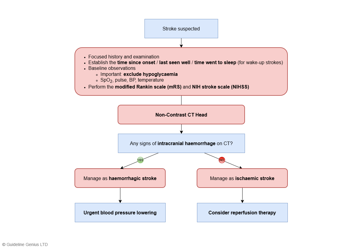
The pathway below illustrates a simplified approach to acute stroke assessment and initial management in the emergency department / hospital setting.
Assessment and Work-Up
Refer all suspected cases of stroke to a hyperacute stroke service.
1. Initial Clinical Assessment
- Exclude hypoglycaemia with a capillary blood glucose test
- Clinical history and assessment, including
- Establish time of onset / last seen well / time went to sleep (for wake-up stroke)
- If possible, perform the NIHSS and mRS
- Check for any thrombolysis contraindications
- Clinical observations (e.g. SpO2, pulse, blood pressure)
- Screen swallowing before the patient receives anything by mouth
2. Brain Imaging
1st line and most important: non-contrast CT head within 1 hour
Radiographic features of intracerebral haemorrhage:
- A hyperdense (bright-white) area of acute blood
- May be surrounded by a hypodense rim of oedema
- +/- Mass effect (e.g. midline shift, ventricular compression)
- Location: often in the basal ganglia, thalamus, posterior fossa
- +/- Intraventricular haemorrhage
Subsequent management:
- If CT shows intracerebral haemorrhage → manage as haemorrhagic stroke (see below)
- If CT shows no haemorrhage → manage as ischaemic stroke (see the Ischaemic Stroke article)
- If the clinical presentation and/or CT suggests subarachnoid haemorrhage → see the Subarachnoid haemorrhage (SAH) article
CT / MR angiogram might be necessary to exclude an underlying macrovascular cause (e.g. aneurysm, AVM)
- Recommended in 18-70 y/o who were previously independent, do not have cancer, and were not taking an anticoagulant (as an underlying macrovascular cause is possible)
- Usually NOT necessary if the patient is >45 y/o with a history of hypertension, and the haemorrhage is located in the basal ganglia, thalamus, or posterior fossa (as in these cases the bleed is almost certainly due to hypertensive small vessel disease)
Management
Acute Management
Note: This section applies to intracerebral haemorrhage (ICH), management of subarachnoid haemorrhage is covered in a separate article – Subarachnoid haemorrhage (SAH)
Patients should be admitted directly to a hyperacute stroke unit for monitoring of their conscious level
The mainstay of acute ICH management focuses on medical stabilisation to prevent the existing bleed from expanding.
| Management aspect / goal | Description |
|---|---|
| Blood pressure lowering | Indications for urgent blood pressure lowering:
Target:
Choice of agent:
Rapid blood pressure lowering should NOT be performed if ANY of the following is present:
|
| Anticoagulant reversal (if appropriate) | Depends on which anticoagulants the patient is on:
|
Surgical interventions:
- Most patients with ICH do NOT require surgical intervention
- Surgery is usually only indicated if there is
- Posterior fossa haemorrhage → haematoma evacuation
- Hydrocephalus (from intraventricular haemorrhage) → EVD insertion
Long-Term Management
Most important: long-term blood pressure control
- Hypertension is the primary treatable risk factor for ICH
- Tight BP control is the most effective way to prevent a recurrent bleed
The more complex aspect is whether to restart antiplatelet or anticoagulant therapy, if the patient was previously taking either
- This should be an individualised decision, made on a case-by-case basis after carefully balancing the risk of recurrent bleeding against the benefits of antiplatelet or anticoagulant therapy
- If antiplatelet therapy is to be restarted, consider at least 24 hours after symptom onset
- For patients with AF who require long-term anticoagulation, left atrial appendage occlusion may be considered as an alternative strategy to reduce the need for long-term anticoagulation
Statins are not routinely used for secondary prevention following ICH itself.
They should only be started where there is a separate cardiovascular indication, in line with the guidance covered in the Lipid Lowering Therapy and Cardiovascular Risk Reduction article.
If a specific underlying cause of ICH is identified, especially a macrovascular abnormality, cause-specific management may be required.
- Aneurysms → endovascular coiling or surgical clipping
- AVMs, dural arteriovenous fistula, cavernous malformations → neurosurgical / interventional radiology management
Microvascular causes, such as hypertensive small vessel disease and cerebral amyloid angiopathy, are mainly managed with strict BP control (see above), rather than a specific procedural intervention.
References
Related Articles
Transient Ischaemic Attack (TIA)