

Basal Cell Carcinoma (BCC)
Basal cell carcinoma (BCC) is the most common type of skin cancer, arising from basal keratinocytes of the epidermis.
This updated UKMLA guide to basal cell carcinoma (BCC) is based on BAD, NICE, and PCDS guidance, which covers risk factors, clinical features, referral criteria, diagnosis and management.
Causes and Risk Factors
BCC is the most common type of skin cancer, accounting for ~70% of all skin cancers.
Key risk factors:
- Cumulative lifetime UV exposure – major risk factor
- Sunlight
- Sunbeds
- Artificial UV exposure (e.g. psoriasis management)
- Fair-skin type (type 1-2)
- Advancing age
- Males
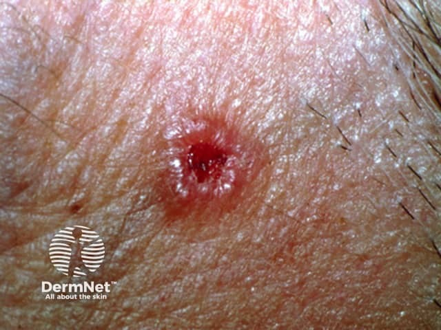
Clinical Features
Typical feature of nodular BCC.
| Location | Most common in the “mask areas” of the face or “H zone”:
|
| Appearance |
|
| Progression |
|
Disclaimer:
This section focuses on the classic features of nodular BCC, which is the most common BCC subtype. Other BCC subtypes exist, including superficial, morphoeic (infiltrative), and mixed BCC, but these are omitted here to keep the article focused on high-yield, non-specialist recognition.
Click to See Clinical Images

Nodular basal cell carcinoma
Source: https://dermnetnz.org/topics/nodular-bcc-images
Nodular basal cell carcinoma
Source: https://dermnetnz.org/topics/nodular-bcc-images
Complications
BCC very rarely metastasises (unlike malignant melanoma and squamous cell carcinoma)
BCC mainly causes local tissue invasion and destruction, particularly on the head and neck
Red Flags and When to Refer
Consider routine referral (to dermatology) if there is a suspected basal cell carcinoma skin lesion.
Since BCC is less dangerous, it only warrants routine referral, while malignant melanoma and squamous cell carcinoma warrant suspected cancer pathway referral.
Investigation and Diagnosis
Dermatologists and other skin cancer specialists can usually diagnose BCC clinically without immediately needing a biopsy
- Thorough skin examination
- Medical photography +/- dermoscopy
Tissue biopsy (most commonly shave or curette biopsy) is NOT strictly mandatory to diagnose BCC, key indications:
- Diagnostic uncertainty
- High-risk type (based on the size and location)
- Suspected recurrence
- To confirm histological subtype (e.g. superficial vs infiltrative), which will impact management
Management
1st line treatment: surgical excision
Surgery-sparing options (e.g. patient unfit for surgery, patient declines surgery)
- Low-risk BCC: topical 5-fluorouracil / imiquimod, cryosurgery, curettage and cautery, photodynamic therapy
- High-risk BCC: radiotherapy (do NOT offer the options for low-risk BCC)
Mohs micrographic surgery may be used for high-risk BCCs (e.g. recurrent lesions or lesions in cosmetically sensitive areas such as the face).
It is a specialised form of surgical excision involving staged removal with immediate microscopic margin assessment.
References
Related Articles
Skin Cancer – Recognition and Referral