

Pre-Malignant Skin Lesions
Pre-malignant skin lesions are abnormal skin lesions with potential to progress into cutaneous malignancy, particularly squamous cell carcinoma. This article covers high-yield pre-mlignant skin lesions relevant to UKMLA-level dermatology based on PCDS guidance, including actinic keratosis, Bowen’s disease, and keratoacanthoma.
Actinic Keratosis (AK)
Definition
AK is a premalignant skin lesion caused by chronic UV exposure, characterised by atypical proliferation of keratinocytes within the epidermis.
The main risk of AK is progression to cutaneous SCC. Although the risk of transformation is low, it increases with time and number of lesions. Having ≥10 AKs is associated with a 14% risk of SCC within 5 years.
Actinic keratosis is also known as solar keratosis.
Causes and Risk Factors
AK arises from cumulative UV exposure
- Those with fair skin, blue eyes and blonde hair are at a higher risk
- In contrast, AK is very rare in those with type 4-6 skin types
- Patients with xeroderma pigmentosum and albinism can develop AK at a very young age
- Artificial UV radiation (e.g. PUVA for psoriasis, sun beds) is also a risk factor
AK is more common in males
Clinical Features
Usually only present in >45 y/o, and the incidence increases with age
Symptoms:
- Usually asymptomatic
Signs:
- Location: areas of sun-exposure (e.g. forearms, hands, head, neck)
- Size: usually <1 cm
- Appearance
- Rough, scaly surface
- White / yellow colour
- Often flat, but the scales can give an elevated appearance
Click to See Clinical Images
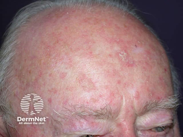
Multiple actinitic keratoses over the forehead
Source: https://dermnetnz.org/topics/actinic-keratosis
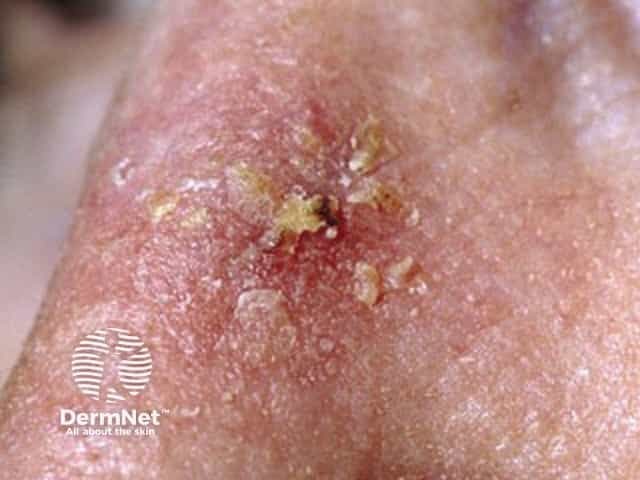
Actinitic keratoses with hyperkeratotic top seen on nasal bridge
Source: https://dermnetnz.org/topics/actinic-keratosis
Management
Referral Criteria
If transformation into SCC is suspected → suspected cancer referral
Red flags:
- Recent growth / change to the AK lesion
- Presence of pain / bleeding / ulceration
- Elevated lesion
PCDS also noted to have a low threshold for referring immunosuppressed patients (esp. post-transplant) as they are at higher risk of developing SCC
Management (Primary Care)
Most AK can be managed in primary care.
| General management |
|
| Pharmacological / interventional management | Up to 25% cases will resolve without treatment, therefore treatment is NOT always necessary (esp. in patients with reduced life expectancy and with small number of lesions)
Treatment options for isolated / small number of lesions:
Treatment options for field change (areas of skin with multiple AKs):
Counselling on 5-FU cream: Patients should be counselled that 5-FU commonly cause an initial inflammatory skin reaction (e.g. erythema, crusting, discomfort) due to destruction of dysplastic keratinocytes The skin may take 6–8 weeks to fully settle. |
References
Bowen’s Disease
Definition
Bowen’s disease is a form of SCC in situ, characterised by full-thickness dysplasia of the epidermis, but WITHOUT invasion through the basement membrane (this distinguishes it from invasive SCC).
Bowen’s disease carries a ~3% risk of transformation into SCC.
Causes and Risk Factors
Bowen’s disease arises from cumulative UV exposure
- Those with fair skin, blue eyes and blond hair are at higher risk
- More common in females
Clinical Features
Symptoms:
- Generally asymptomatic
Signs:
- Lesions can be single or multiple
- Very slow-growing
- Location: sun-exposed sites (e.g. forearms, hands, head, neck) (esp. lower legs in females)
- Colour: pink
- Well-defined, pink and scaly patches or plaques (flat)
- As lesions grow they may become crusty, fissured or ulcerated
Bowen’s disease tends to have finer scale, less substance and more sharply demarcated borders than actinic keratosis, helping differentiate the two clinically.
Click to See Clinical Images
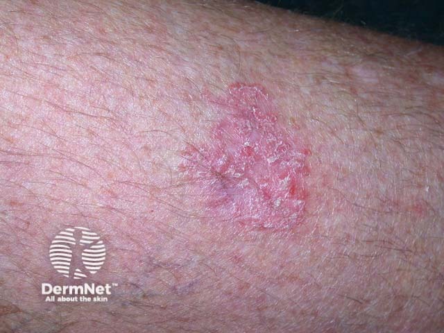
Bowen’s disease
Source: https://dermnetnz.org/topics/intraepidermal-carcinoma-images
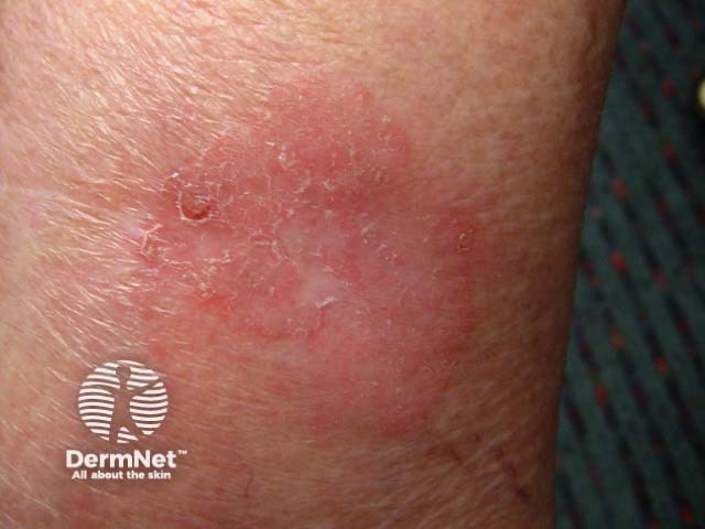
Bowen’s disease
Source: https://dermnetnz.org/topics/intraepidermal-carcinoma-images
Management
Perform a thorough skin examination to look for cutaneous malignancies (e.g. melanoma)
1st line treatment:
- Cryotherapy (but avoid in the gaiter area of the leg and other areas of poor skin healing)
- 5-Fluorouracil cream (5-FU)
Counselling on 5-FU cream:
Patients should be counselled that 5-FU commonly cause an initial inflammatory skin reaction (e.g. erythema, crusting, discomfort) due to destruction of dysplastic keratinocytes
The skin may take 6–8 weeks to fully settle.
References
Keratoacanthoma
Definition
Keratoacanthoma is a rapidly evolving tumour of the skin, composed of keratinising squamous cell originating in pilosebaceous follicles.
It is thought to represent a well-differentiated variant of SCC.
Clinical Features
Sun exposure plays a role. Some cases keratoacanthoma arise following an injury to the skin.
Keratoacanthoma is rapid growing and evolving:
- Early: small, firm, rounded, skin-coloured to red papule
- Late (“volcano-like appearance“): symmetrical, well-defined dome-shaped nodule with a central keratin core
- The lesion characteristically resolve after ~3 months
Click to See Clinical Images
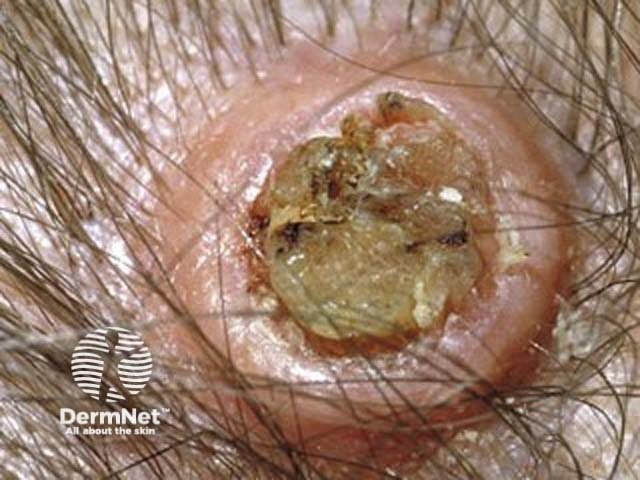
Keratoacanthoma
Source: https://dermnetnz.org/topics/keratoacanthoma-images
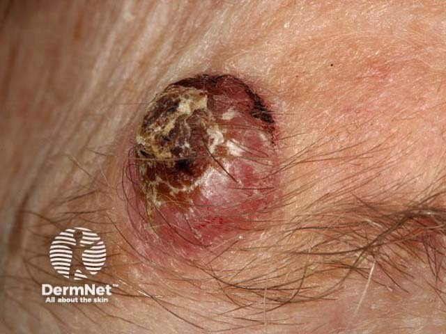
Keratoacanthoma
Source: https://dermnetnz.org/topics/keratoacanthoma-images
Management
ALL cases should be referred urgently (suspected cancer pathway) to secondary care
- Rationale: It is difficult to distinguish between a keratoacanthoma and SCC
Keratoacanthoma should be managed as SCC in secondary care (i.e. surgical excision where possible) – see the Cutaneous Squamous Cell Carcinoma (cSCC) article for more information.
References
Related Articles
Skin Cancer – Recognition and Referral