Assessment and management of VTE in pregnancy is covered in a separate article: Venous Thromboembolism (VTE) During Pregnancy
Background information
Definition
Venous thromboembolism (VTE): is a term that encompasses deep vein thrombosis (DVT) and pulmonary embolism (PE).
- Deep vein thrombosis: clot formation (thrombosis) within a deep vein; most commonly of the legs or pelvis.
- Pulmonary embolism: life threatening condition characterised by the presence of emboli, usually arising from a DVT, in the pulmonary arterial system.
Provoked DVT/PE (~2/3 of cases): occuring in the presence of a recent (within 3 months) major clinical risk factor for VTE
Unprovoked DVT/PE (~1/3 of cases): occuring in the absence of a recent (within 3 months) major clinical risk factor for VTE
Whether VTE is provoked/unprovoked plays a role in the duration of anticoagulation on NICE guidelines
Epidemiology
- Common; VTE is the third most common cardiovascular disease (after acute myocardial infarction & stroke)
- VTE cases
- 1/3 → PE
- 2/3 → DVT
- Incidence increases sharply with age / presence of risk factors
- VTE cases
Aetiology
Deep Vein Thrombosis
DVT is caused by risk factors that fall under Virchow’s triad
Virchow’s triad encompasses the 3 major mechanisms that cause thrombosis
- Hypercoagulability
- Endothelial / vessel wall damage
- Venous stasis
Risk factors for DVT grouped by Virchow’s triad
1. Hypercoagulability
- Personal / family history of VTE
- Active cancer
- Pregnancy / Puerperium
- Hormone therapy (COCP / HRT)
- APS / thrombophilias (familial/acquired)
- Inflammatory, pro-thrombotic disorders
- Diabetes mellitus
- Smoking
- Male sex (particularly for DVT)
- Dehydration
2. Endothelial damage
- Recent trauma or lower limb fracture
- Surgery (direct vessel trauma) – esp major surgeries
- Direct venous trauma (e.g. IV cannula, indwelling central venous catheter)
- Recent myocardial infarction (< 3 months)
- Varicose veins / Superficial venous thrombosis
3. Venous stasis
- Significant immobility / hospitalisation / bed rest >5 days
- Prolonged travel (> 4 hours)
- Heart failure
- Obesity (BMI >30)
- Increasing age (↑ incidence / mortality)
Pulmonary Embolism
Most common cause → DVT
Other → causes of non-thrombotic embolism:
- Fat embolism
- Air embolism
- Amniotic fluid embolism
- Septic/Bacterial embolism
Clinical features
Deep Vein Thrombosis
Localisation → DVT is typically unilateral
Symptoms
- Leg pain / tenderness
- Throbbing, cramping or dull ache
- Worse on walking/weight bearing
- Swelling (often calf or thigh)
- Feeling of tightness or heaviness
- Systemic features
- Low-grade fever, malaise
- If concurrent → symptoms of pulmonary embolism (e.g., dyspnoea, chest pain)
Signs / Examination Findings
- Leg swelling
- Calf-circumference difference
- Pitting oedema
- Erythema → redness, warmth or livid discoloration
- Dilated superficial veins
- Local tenderness on palpation
- Special tests (less reliable, non-diagnostic but often mentioned)
- Homans sign → calf pain on foot dorsiflexion
- Meyer sign (calf-squeeze) → calf pain on compression
Pulmonary Embolism
Symptoms
- Onset → typically sudden
- Dyspnoea → most common symptom (~50% of cases)
- Typically persistent or progressive
- Pleuritic chest pain (~40% of cases) OR retrosternal chest pain (less common)
- Cough ± haemoptysis
- Syncope or presyncope → more in massive PE
- Systemic features (fever, dizziness, weakness)
- Symptoms of DVT may be present
Signs / Examination findings
- General comments
- Signs / Examination findings are often nonspecific.
- Many patients have a normal physical examination (esp in smaller emboli)
- Potential findings
- Observations
- Tachypnoea / Tachycardia (common)
- Hypoxia
- Fever (uncommon, typically low-grade)
- Hypotension / shock → suggests massive PE
- Right heart strain
- Elevated JVP
- Heart auscultation → loud P2, widely split S2
- Pleural rub
- Observations
Complications
Deep Vein Thrombosis
- Pulmonary embolism
- Recurrent DVT
- Post-thrombotic syndrome (PTS) → up to 50% within 2 years of lower limb DVT
- Definition → chronic venous insufficiency in the affected limb, secondary to DVT
- Clinical Dx → symptoms of chronic venous insufficiency (e.g., limp pain, swelling, oedema, skin hyperpigmentation), typically, 3-6 months after initial DVT event
Pulmonary Embolism
Acute
- Arrhythmias
- Respiratory failure
- Right ventricular failure ± haemodynamic instability
- Sudden cardiac death (often due to PEA)
Chronic
- Chronic thromboembolic pulmonary hypertension (CTEPH)
- Rare & severe complication (progresses to right heart failure)
- Subtype of pulmonary hypertension caused by chronic thromboembolic occlusion of pulmonary vessels
Deep Vein Thrombosis Guidelines
Assessment and Management Algorithm
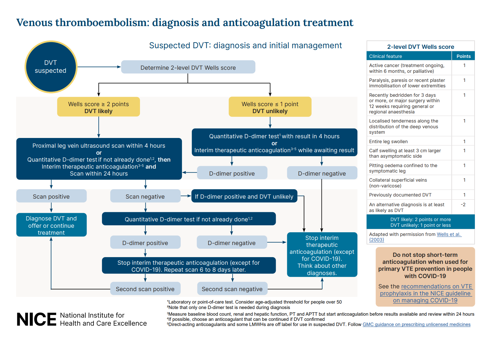
If DVT is suspected, calculate the two-level DVT Wells score
| Clinical feature | Points |
|---|---|
| Active cancer (or within 6 months) | 1 |
| Lower limb immobilisation (recent plaster use, paralysis, paresis) | 1 |
| Recently bedridden for ≥3 days or major surgery within 12 weeks requiring general or regional anaesthesia | 1 |
| Localised tenderness along the distribution of deep venous system | 1 |
| Entire leg swollen | 1 |
| Calf swelling >3 cm larger than the other leg | 1 |
| Pitting oedema confined to the affected leg | 1 |
| Collateral superficial veins (non-varicose) | 1 |
| Previously documented DVT | 1 |
| An alternative diagnosis is at least as likely as DVT | -2 |
Interpretation:
- Score 2 or more: DVT likely
- Score 1 or less: DVT unlikely
DVT Likely (Wells 2 or more)
Perform a proximal leg vein ultrasound (with results available within 4 hours)
If the ultrasound cannot be done within 4 hours, perform the following:
- Perform D-dimer test, and
- Offer interim therapeutic anticoagulation, and
- Ensure ultrasound is done within 24 hours
Abnormal Ultrasound
Diagnose DVT and start treatment
Normal Ultrasound
Perform D-dimer test
- -ve D-dimer → DVT unlikely, consider alternative diagnosis (stop any interim anticoagulation)
- +ve D-dimer → repeat proximal leg vein ultrasound 6-8 days later (stop any interim anticoagulation)
- Abnormal ultrasound → start treatment
- Normal ultrasound → consider alternative diagnosis
A single negative proximal ultrasound does not definitively exclude DVT in patients with high clinical suspicion or elevated D-dimer.
Anticoagulation (e.g., apixaban) should not be continued unless DVT is confirmed, due to bleeding risk and lack of proven benefit in the absence of objective evidence of thrombosis
DVT Unlikely (Wells 1 or less)
Perform D-dimer test (with results available within 4 hours):
- -ve D-dimer → DVT unlikely, consider alternative diagnosis (stop any interim anticoagulation)
- +ve D-dimer → perform a proximal leg vein ultrasound (with results available within 4 hours)
- If ultrasound not possible → offer interim therapeutic anticoagulation and ensure ultrasound is done within 24 hours
If D-dimer cannot be done within 4 hours → offer interim therapeutic anticoagulation while waiting
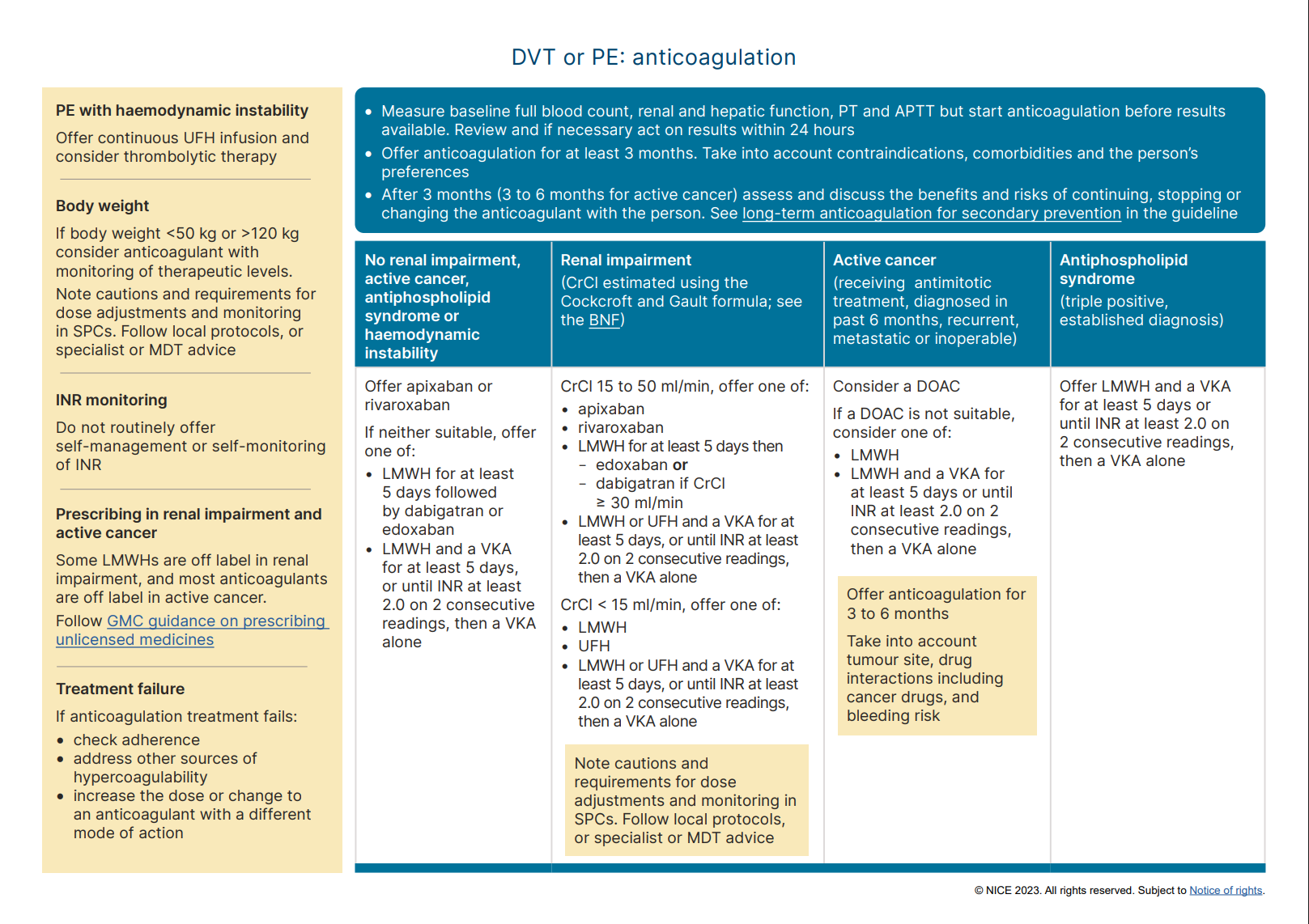
Treatment For Confirmed DVT
This section is identical for DVT and PE
The drug class of choice to treat DVT and PE are anticoagulants.
Choice of Drug
1st line:
- Apixaban or rivaroxaban
2nd line:
- Warfarin with LMWH lead-in, or
- Dabigatran or edoxaban with LMWH lead-in
In renal impairment (not renal failure), apixaban is preferred over rivaroxaban as it has less renal excretion.
Choice of Drug in Specific Patient Populations
| Patient population | Recommended drug |
|---|---|
| Renal failure (creatinine clearance <15 mL/min) | Avoid DOAC, use:
|
| Active cancer |
|
| Pregnancy | DOAC and warfarin are contraindicated, use:
|
| Anticoagulation contraindicated | Consider inferior vena cava filter |
| Antiphospholipid syndrome (triple positive) | Warfarin with LMWH lead-in |
Duration of Treatment
- Provoked: 3 months
- Unprovoked: 6 months
- Concurrent cancer: 3-6 months
Pulmonary Embolism (PE) Guidelines
Assessment and Management Algorithm
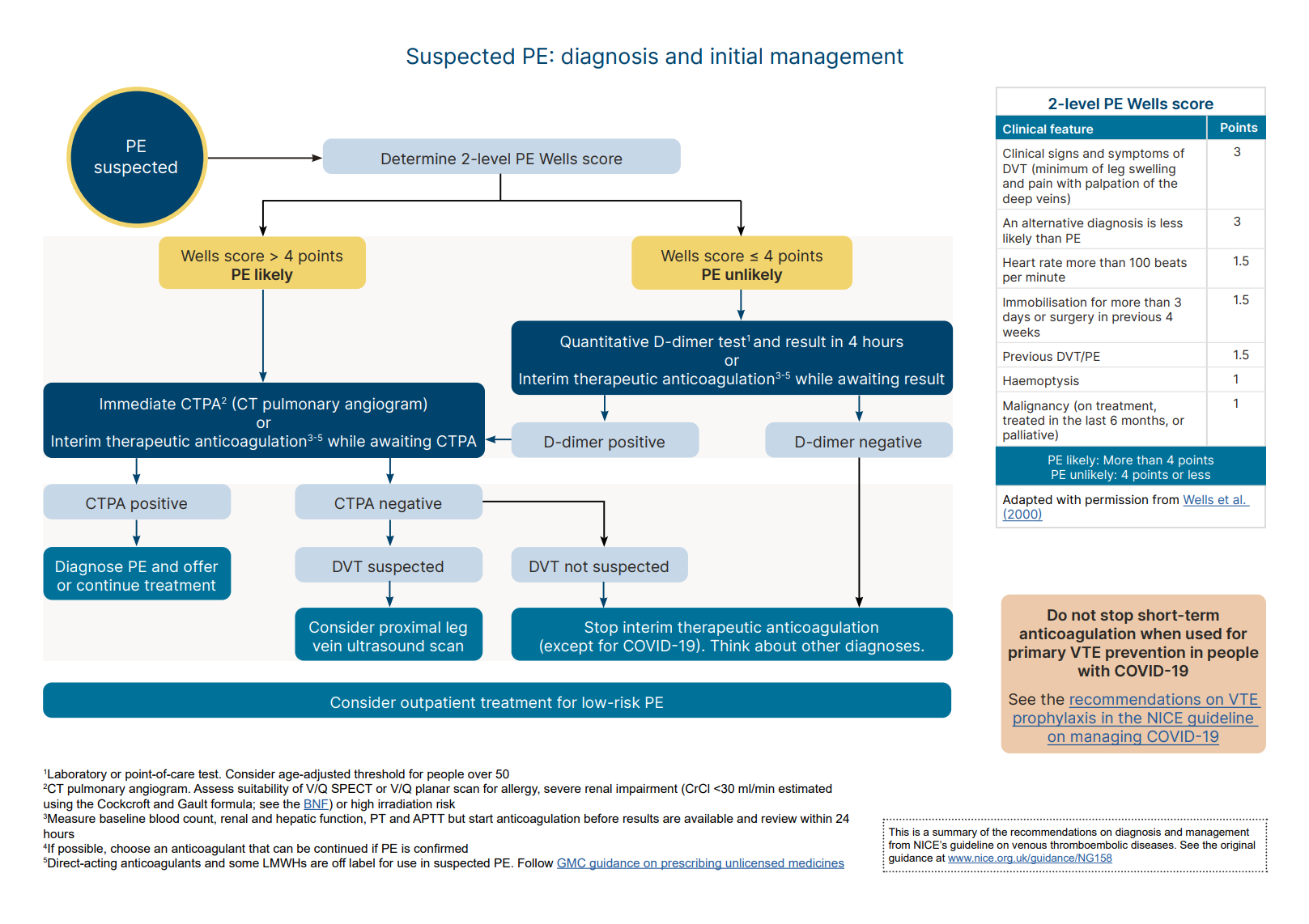
Initial steps:
- Perform a chest X-ray to exclude other causes (along with general history and physical examination)
- If the clinical suspicion is low based on history, examination and initial investigations (e.g. X-ray) and alternative diagnoses are feasible → consider using the PERC to help determine whether further investigations for PE are necessary
If PE is suspected, calculate the two-level PE Wells score to guide subsequent steps:
| Clinical feature | Points |
|---|---|
| Clinical features of DVT (minimum of leg swelling and pain with palpation of the deep veins) | 3 |
| An alternative diagnosis is less likely than PE | 3 |
| Heart rate >100 bpm | 1.5 |
| Immobilisation for >3 days or surgery in the previous 4 weeks | 1.5 |
| Previous DVT / PE | 1.5 |
| Haemoptysis | 1 |
| Active cancer (or within 6 months) | 1 |
Interpretation:
- Score 5 or more: PE likely
- Score 4 or less: PE unlikely
PE Likely (Wells 5 or more)
Perform a CT pulmonary angiogram (CTPA) immediately
- Abnormal CTPA → diagnose PE and start treatment
- Normal CTPA
- If DVT is suspected → consider a proximal leg vein ultrasound scan
- If DVT is not suspected → PE unlikely, consider alternative diagnosis (stop any interim anticoagulation)
If CTPA is not appropriate (allergic to contrast or severe renal impairment or high risk from irradiation) → consider V/Q SPECT or planar scan as an alternative
If CTPA / V/Q scan cannot be done immediately → offer interim therapeutic anticoagulation while awaiting CTPA
Bedside echocardiography to assess right ventricular strain is an appropriate alternative in those who are not suitable candidates for CTPA (e.g. clinically unstable for imaging, allergic to contrast, severe renal impairment, high risk from irradiation).
Note that this is not mentioned in NICE guidelines but is commonly performed in practice, and endorsed by international echocardiography guidelines.
PE Unlikely (Wells 4 or less)
Perform D-dimer test (with results available within 4 hours):
- -ve D-dimer → PE unlikely, consider alternative diagnosis (stop any interim anticoagulation)
- +ve D-dimer → perform CTPA (or V/Q scan) immediately (i.e. follow the above PE likely algorithm)
If D-dimer cannot be done within 4 hours → offer interim therapeutic anticoagulation while waiting
Bedside echocardiography to assess right ventricular strain is an appropriate alternative in those who are not suitable candidates for CTPA (e.g. clinically unstable for imaging, allergic to contrast, severe renal impairment, high risk from irradiation).
Note that this is not mentioned in NICE guidelines but is commonly performed in practice, and endorsed by international echocardiography guidelines.
PE with Haemodynamic Instability
Hemodynamic instability is indicated by any of the following: [Ref]
- Hypotension (SBP <90 mmHg, or >40 mmHg drop that are not explained by other causes)
- End-organ hypoperfusion (features inc. altered mental status, cold/clammy skin, oliguria, or elevated lactate)
- Cardiac arrest
This clinical scenario is also termed “high-risk” or “massive” pulmonary embolism
Offer Immediate Reperfusion Therapy:
- Continuous UFH infusion, and
- Consider thrombolytic therapy (e.g. alteplase)
The presence of right ventricular dysfunction (confirmed on echocardiography & supported by elevated cardiac biomarkers) is NOT an indication for thrombolysis by itself (unless there is hemodynamic instability).
Treatment For Confirmed PE
The drug class of choice to treat DVT and PE are anticoagulants, NOT antiplatelets (e.g. aspirin, clopidogrel).
For pulmonary embolism, outpatient treatment may be appropriate for low-risk PE
- NICE recommends using a validated tool (pulmonary embolism severity index is an example, but NICE did not recommend any specific tool)
- Management principles are the same as following with additional patient education and safety netting
- Rationale: no evidence showed that outpatient treatment is less effective or less safe than inpatient treatment for people with low-risk PE
The section below is identical for DVT and PE.
Choice of Drug
1st line:
- Apixaban or rivaroxaban
2nd line:
- Warfarin with LMWH lead in, or
- Dabigatran or edoxaban with LMWH lead in
Choice of Drug in Specific Patient Populations
| Patient population | Recommended drug |
|---|---|
| Renal failure (creatinine clearance <15 mL/min) | Avoid DOAC, use:
|
| Active cancer |
|
| Pregnancy | DOAC and warfarin are contraindicated, use:
|
| Anticoagulation contraindicated, or PE happened while on anticoagulation | Consider inferior vena cava filter |
| Antiphospholipid syndrome (triple positive) | Warfarin with LMWH lead in |
Duration of Treatment
- Provoked: 3 months
- Unprovoked: 6 months
- Concurrent cancer: 3-6 months
References
NICE Algorithm – DVT Diagnosis and Initial Management
NICE Algorithm – PE Diagnosis and Initial Management
NICE Visual Aid – Anticoagulation in DVT and PE