Background Information
Definitions and Terms
| Term | Definition | Notes |
|---|---|---|
| Ocular hypertension | Raised IOP (>21 mmHg) WITHOUT optic nerve damage / visual field loss | At risk of developing glaucoma |
| Glaucoma | Chronic optic nerve neuropathy characterised by optic disc cupping and visual-field defects
Usually but NOT always associated with ocular hypertension |
Umbrella term that includes:
|
| Normal tension glaucoma | Chronic optic nerve neuropathy with NORMAL IOP | Thought to involve vascular or structural susceptibility factors rather than IOP alone |
| Chronic open-angle glaucoma | Characterised by:
|
The most common form of glaucoma
Most cases present chronically |
| Angle-closure glaucoma | Characterised by raised IOP secondary to a closed anterior chamber angle | Most cases present acutely |
Raised IOP ≠ glaucoma. Optic nerve neuropathy is needed for it to be glaucoma, indicated by optic disc cupping and visual-field defects.
Guidelines
Investigation and Diagnosis
Tests in Primary Care (before referral)
Offer ALL the following tests before referral:
- Central visual field assessment using automated perimetry
- IOP measurement with tonometry (e.g., measured with Goldmann aplanation tonometer)
- Optic nerve assessment and fundus examination with stereoscopic slit lamp biomicroscopy and OCT or optic nerve head image
- Peripheral anterior chamber configuration and depth assessment with gonioscopy / van Herick test / OCT
After the above tests, refer if ANY of the following:
- IOP ≥24 mmHg
- Visual field defect
- Optic nerve head damage
Tests in Secondary Care
Offer ALL the following tests to diagnose COAG:
- Central visual field assessment using automated perimetry
- IOP measurement with Golmann-type applanation tonometry (slit lamp mounted)
- Optic nerve assessment and fundus examination with stereoscopic slit lamp biomicroscopy with pupil dilatation
- Peripheral anterior chamber configuration and depth assessment with gonioscopy
- Central corneal thickness measurement
- A thin cornea underestimates true IOP, and a thick cornea overestimates IOP
Management
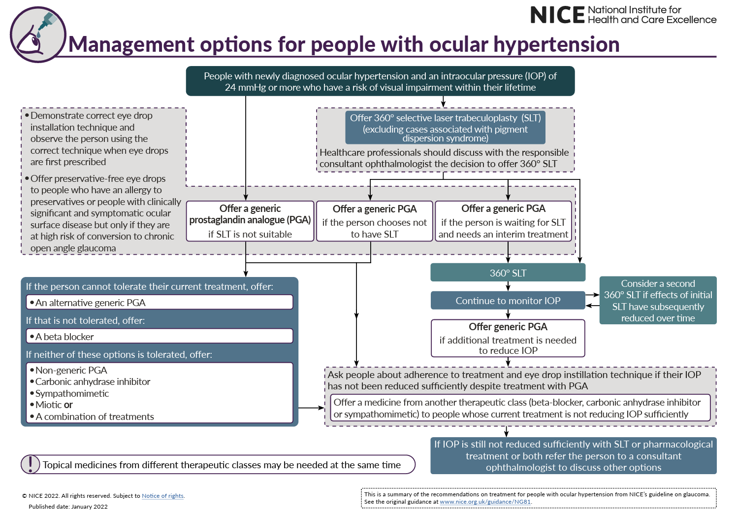
Ocular Hypertension – Management
Indications to treat: IOP ≥24 mmHg + at risk of visual impairment within their lifetime
- 1st line: 360° selective laser trabeculoplasty (SLT)
- Consider a second attempt if the initial successful SLT has reduced over time
- 2nd line: prostaglandin analogue eye drops
- 3rd line: beta blocker eye drops
- 4th line: beta blocker / carbonic anhydrase inhibitor / sympathomimetic eye drops
360° SLT can delay the need for regular use of topical eye drops, but they will still be necessary at some point.
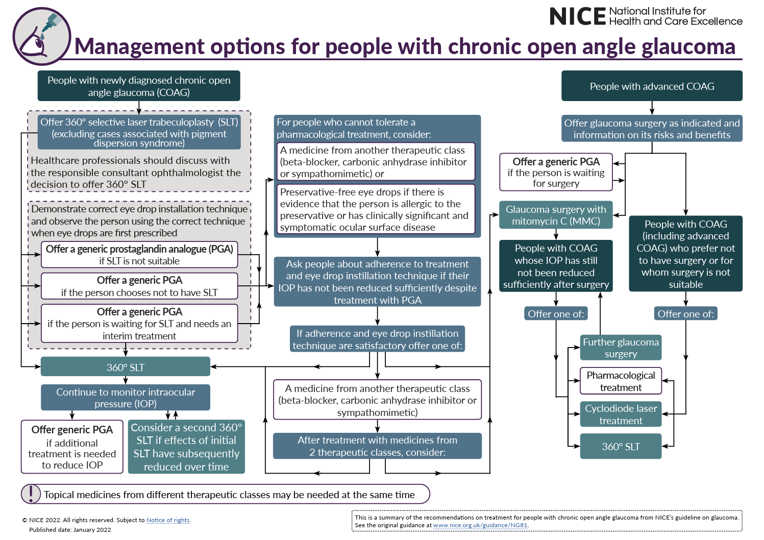
COAG – Management
Indications to treat: IOP ≥24 mmHg OR at risk of visual impairment within their lifetime
Non-Advanced COAG – Management
- 1st line: 360° selective laser trabeculoplasty (SLT)
- Consider a second attempt if the initial successful SLT has reduced over time
- 2nd line: prostaglandin analogue eye drops
- 3rd line: beta blocker / carbonic anhydrase inhibitor / sympathomimetic eye drops OR glaucoma surgery (e.g., surgical trabeculectomy) with mitomycin-C
- 4th line: cyclodiode laser treatment
360° SLT can delay the need for regular use of topical eye drops, but they will still be necessary at some point.
Advanced COAG – Management
- Glaucoma surgery (e.g., surgical trabeculectomy), AND
- Pharmacological augmentation with mitomycin-C
Monitoring and Reassessment
At each assessment, offer the Goldmann applanation tonometry (slit lamp mounted)
If clinically indicated, also offer:
- Anterior segment slit lamp examination with van Herick peripheral anterior chamber depth assessment
- Repeat gonioscopy
- Repeat visual field testing with automated perimetry
- Repeat assessment of the optic nerve head
References
NICE Visual Aid – Management of Ocular Hypertension
NICE Visual Aid – Management of Chronic Open Angle Glaucoma